Uterine fibroids
Uterine fibroids are one of the most common benign tumors in women of childbearing age. They occur in 20–40% of women, but their actual prevalence may be higher, since the disease often proceeds without pronounced symptoms and remains unnoticed. In addition, there is a tendency for the pathology to become “rejuvenated”: fibroids are increasingly detected in women under 30, and often in forms that require surgical intervention. This diagnosis can cause anxiety, especially if it is made for the first time.

specialists

equipment

treatment
Uterine fibroids signs and symptoms
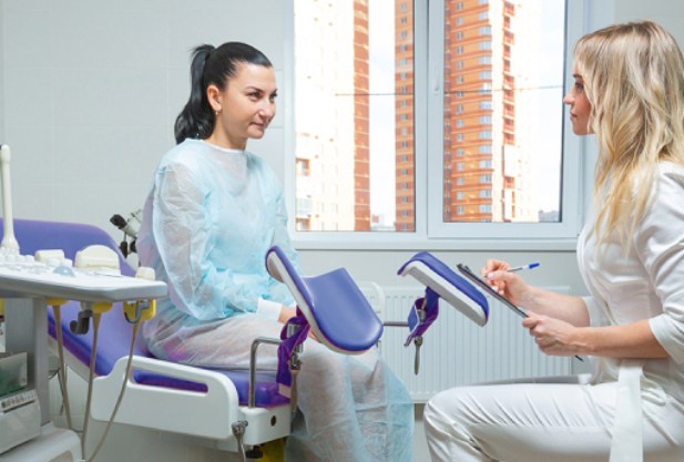
Symptoms of uterine fibroids depend on the location of the formation. Subserous nodes can compress nearby organs (ureters, bladder, rectum), which can cause heaviness in the lower abdomen and pain, as well as dysfunction of the bladder and intestines, including constipation, painful or involuntary urination. Perhaps the appearance of pain in the vagina during intercourse, discomfort in the lower back - these symptoms are less common and are often associated with other problems with women's health.
Submucous and interstitial fibroids are characterized by heavy, prolonged menstruation, increasing the risk of uterine bleeding, there may be bloody discharge in the intermenstrual period. Therefore, women with uterine fibroids often develop anemia, which is accompanied by weakness, fatigue, dizziness.
Small interstitial fibroids do not cause discomfort, so the pathology is asymptomatic for a long time. As soon as the tumor begins to increase in size, the first main signs appear.
Types of operations to remove uterine fibroids
Laparoscopic myomectomy
It is performed through small punctures in the abdominal wall using a laparoscope, an instrument with a camera and surgical attachments. The method minimizes trauma to surrounding tissues, which facilitates rapid patient recovery. The procedure takes 1.5–2 hours, and hospitalization usually lasts no more than 1–2 days.
Hysteroscopic myomectomy
The procedure is designed to remove fibroids that grow into the uterine cavity (submucosal fibroids). It is performed through the vagina using a hysteroscope, eliminating the need for skin incisions. The method is suitable for small fibroids and allows preserving the integrity of the uterus.
Laparotomic myomectomy
It is used to remove large fibroids or in cases of multiple lesions. The procedure is performed through an abdominal incision, providing access to hard-to-reach areas. This method requires a longer recovery period (4–6 weeks) and is applied when laparoscopy or hysteroscopy is ineffective.
Hysterectomy
This is a radical surgery involving the complete removal of the uterus. It is prescribed for severe complications, such as heavy bleeding or suspected malignant transformation. There are several techniques: through the abdominal wall, vagina, or using laparoscopic equipment to reduce trauma.
Uterine artery embolization (UAE)
(UAE) is a minimally invasive procedure aimed at blocking the blood supply to fibroids, leading to their reduction.
How is the procedure performed? The patient is under local anesthesia. Through a small puncture in the thigh, the doctor inserts a catheter into the uterine arteries and delivers micro-particles that block the blood supply to the fibroids. The procedure takes about 30–60 minutes.
What happens after UAE?
- The fibroids begin to shrink in size, and symptoms (pain, bleeding) disappear within a few months.
- Recovery usually takes 7–10 days, after which the woman can return to her normal lifestyle.
However, this method is not suitable for suspected malignancy or cases where fibroids are outside the vascular zone.
FUS ablation
A modern technique for destroying uterine fibroids using focused ultrasound under MRI control. The method does not require incisions, making it attractive for patients with single fibroids who do not plan pregnancy.
Radiofrequency ablation (RFA)
A minimally invasive procedure that uses radio waves to destroy fibroids. It is suitable for patients with moderate symptoms who prefer a gentle and less traumatic approach to treatment.
Causes of the disease and treatment of uterine fibroids
"Uterine fibroids are formed mainly against the background of hormonal imbalance associated with a violation of the level of estrogens and progesterone - key female hormones. This imbalance stimulates abnormal cell growth in the muscular layer of the uterus, which is called the myometrium. However, hormones are not the only reason: there are other predisposing factors, such as genetic predisposition, chronic inflammation and even lifestyle," explains the gynecologist at the K+31 clinic.
It is worth noting that even in the absence of obvious risk factors, uterine fibroids can develop spontaneously.
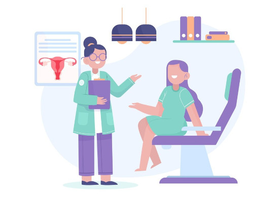
Main causes and risk factors:
- Heredity If close relatives on the mother's side have been diagnosed with fibroids, the likelihood of its development increases significantly
- Hormonal disruptions Early onset of menstruation, late onset of menopause, cycle irregularities, as well as the absence of pregnancy and childbirth - all this creates conditions for the formation of uterine fibroids
- Excess weight Adipose tissue is an additional source of estrogen, which can accelerate the development of a tumor
- Constant stress Emotional overload can disrupt the functioning of the hormonal system, increasing the risk of uterine nodes
- Endocrine diseases Problems with the thyroid gland, adrenal glands or other endocrine organs often affect reproductive health
- Environmental factors Environmental pollution, exposure to toxins and xenoestrogens (chemical compounds that imitate the action of estrogens) negatively affect the tissues of the uterus
- Poor nutrition A diet with excess fat and sugar and a lack of fiber, vitamins and microelements can lead to hormonal imbalances
- Low physical activity A sedentary lifestyle slows down metabolism and has a negative impact on the endocrine system
Answers to popular questions
Doctors' answers:
Can fibroids go away on their own?
Is it possible to get pregnant with uterine fibroids?
How fast does fibroid grow?
Is it necessary to remove the fibroid?
What are the restrictions associated with fibroids?

This award is given to clinics with the highest ratings according to user ratings, a large number of requests from this site, and in the absence of critical violations.

This award is given to clinics with the highest ratings according to user ratings. It means that the place is known, loved, and definitely worth visiting.

The ProDoctors portal collected 500 thousand reviews, compiled a rating of doctors based on them and awarded the best. We are proud that our doctors are among those awarded.
Make an appointment at a convenient time on the nearest date
Price
Other services
Hormone therapy
Radio wave gynecology with the Surgitron deviceLaser therapy using the Photona device
Sling operations Ectopic pregnancy Delayed menstruation Removal of the uterus (hysterectomy) Thrush (vaginal candidiasis) Prolapse of the uterus and vagina Uterine polyp (endometrial polyp) Cervical dysplasia Adenomyosis Treatment of sexual infections Vaginitis (Colpitis) Erythroplakia of the cervix Endometritis Bacterial vaginosis Symphysitis (symphysiopathy) Erosion and ectopia of the cervix Vulvovaginitis Premenopause Uterine artery embolization for uterine fibroids Cervicitis Gynecologist consultation Dysmenorrhea (painful periods) Amenorrhea Removal of the ovaries (oophorectomy) Postmenopausal Sphinctermetry Treatment and intimate rejuvenation with the Fotona laser Adenomyosis (Endometriosis of the uterus) Vulvitis Vaginal surgeries Inflammation of the appendages (adnexitis, salpingo-oophoritis) Labiaplasty (labiaplasty) Bartholinitis Surgery to remove an ovarian cyst Prolapse (prolapse) of the uterus and vagina Hormone replacement therapy (HRT) First menstruation 7 days after embryo transfer Biochemical pregnancy IVF protein diet Day 5 after embryo transfer Follicles Bicornuate uterus and pregnancy Day 9 after embryo transfer 1 day after embryo transfer Age and Fertility 10 days after embryo transfer



















About the disease
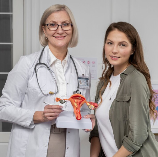
Predisposing factors to the development of fibroids are damage to the uterus due to abortion, surgical interventions, hormonal disorders, including after pregnancy, concomitant gynecological diseases, including inflammatory processes, endometriosis, cysts, etc. A special role in this issue is played by genetic predisposition. Fibroids can develop at any age.
By localization, fibroids are divided into interstitial (located in the wall of the uterus), subserous (growing outside the uterus), submucous fibroids (growing towards the uterine cavity).
Answering the question of what uterine fibroids are, it is important to note that myomatous nodes can be single or multiple.
To prevent further growth of uterine fibroids and avoid surgery, a woman needs to contact a specialist as soon as possible, who will prescribe her effective treatment with hormones and other drugs.