Modern radiation diagnostics uses a large number of research methods that make it possible to diagnose more than two thousand different diseases. Along with magnetic resonance imaging, ultrasound and radionuclide diagnostics, the x-ray method of studying the internal organs of a person makes it possible to establish or supplement about 80 percent of all clinical diagnoses.
X-ray of the bones and joints of the extremities, which you can undergo at the K+31 clinic in Moscow, is a safe and painless procedure that allows you to timely diagnose pathologies of the osteoarticular system and identify complications or secondary manifestations.
Having a gentle effect on human organs and tissues, the procedure of modern radiological examination is virtually harmless to the patient, in addition, it is one of the fastest and most inexpensive. If high-quality X-rays are available, the doctor can identify pathologies in the initial stages, determine the exact location of the injury, and damage to internal organs.
To conduct an X-ray examination of a patient, virtually no preparation for the procedure is required. The subject only needs to free the body area from clothing and other objects. The list of contraindications for this type of diagnosis is also very small. It is not recommended to undergo procedures involving X-ray radiation for women during pregnancy, as well as for young children unless absolutely necessary.
All types of X-ray examinations are carried out with maximum comfort for the patient. The availability of modern programs helps to minimize the patient’s radiation dose during radiography and fluoroscopy. And advanced X-ray image processing capabilities allow you to avoid taking additional targeted images.
Immediately put the patient in the right place and do not redo the low-quality image, i.e. do not re-irradiate the patient
Use extremely low doses of radiation to obtain high-quality images
Take as many pictures as the doctor prescribes
Plain radiography makes it possible to obtain a picture of the entire area or organ under study. When prescribing two x-rays per day, the doctor must take into account the total radiation exposure.
K+31 conducts all types of examinations of the chest organs, including radiography in 2 projections, as well as the gastrointestinal tract, lungs, genitourinary and osteoarticular systems. It is possible to supplement diagnostic and therapeutic endoscopic procedures under the control of fluoroscopy, as well as fistulography.
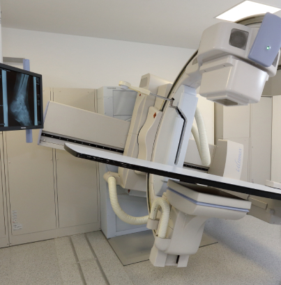
Universal teleoperated turntable-tripod system that provides unhindered access and automated patient positioning without X-ray centering.
- Automatic image stitching and digital subtraction.
- Single-sided support and adjustable table top height
- Rotating X-ray tube allows for diagnostics in patients with limited mobility.
- To reduce radiation exposure, collimation and positioning of the patient are performed without X-ray radiation.
- Continuous and pulsed fluoroscopy at up to 15 frames per second.
If you are wondering where to take an x-ray of internal organs, bones or joints, please contact the radiology department of the K+31 clinic. Qualified specialists conduct all types of research on the most modern equipment. The website of the Center contains information about services, prices are indicated in rubles.
Price
Our services

This award is given to clinics with the highest ratings according to user ratings, a large number of requests from this site, and in the absence of critical violations.

This award is given to clinics with the highest ratings according to user ratings. It means that the place is known, loved, and definitely worth visiting.

The ProDoctors portal collected 500 thousand reviews, compiled a rating of doctors based on them and awarded the best. We are proud that our doctors are among those awarded.
Answers to popular questions
What is radiography?
X-ray is a method for diagnosing internal organs, muscle tissue and bones. During the examination, an X-ray is taken of a specific organ to identify diseases, and the patient is given a photograph with visualization. With radiography, which is carried out over a longer period of time, the image can be obtained on:
- Film (direct or analogue radiography)
- Digital device, disk (digital X-ray)
How does X-ray work?
In an X-ray machine, X-rays are generated in a tube, which are absorbed differently by different tissues of the body. Bones absorb rays best, so they appear white on an X-ray. The darkest areas are muscle tissue, fluids, and air that fills the lung cavities.
The radiologist interprets the x-rays and explains what the x-ray shows. The images are then sent to your attending physician.
How does radiography differ from fluoroscopy?
During radiography, a specialist takes x-rays simultaneously, which allows you to obtain an image of the internal organs and assess their condition at the time of fixation. With fluoroscopy, images are taken in real time while the organ is functioning.
Fluoroscopy (x-ray) is an advanced diagnostic method. But during this procedure the patient receives a large dose of radiation.
How often can an x-ray be taken?
Any x-ray examination always means that the patient receives a dose of radiation. Therefore, it is recommended to do x-rays or fluoroscopy no more than once a year. According to indications, treatment is monitored 2 times a year.
Who interprets radiography?
What types of x-ray examinations are carried out at K+31?
- X-ray of the skull
- X-ray of the cervical spine
- X-ray of the hip joint
- Chest X-ray
- X-ray of the lumbosacral region
- X-ray of the paranasal sinuses
- X-ray of the wrist joint
- X-ray of the knee joint
- X-ray of the abdominal cavity
- Intestinal X-ray
- X-ray of the ankle
- Urography
- Mammography
- Pelvic x-ray
- X-ray of thighs
- X-ray of the heel bone
- X-ray of the lower leg
- X-ray of the duodenum
- X-ray of the esophagus
- X-ray of the collarbone
- Digital radiography
- X-ray of the foot
- X-ray of the stomach
- Breast X-ray
- Fluorography
- X-ray of the femur
- X-ray of the tibia
- X-ray of the coccyx
- X-ray of the nasopharynx
- Kidney X-ray
- X-ray of the sella turcica
- X-ray of the humerus
- X-ray of the lumbosacral region
- X-ray of the paranasal sinuses
- X-ray of the skull bones
- X-ray of the nasal bones
- X-ray of bone structures
- X-ray of soft tissues
Make an appointment at a convenient time on the nearest date


Based on varying degrees of absorption of X-rays by human tissue, the X-ray diagnostic method has long and firmly taken its place among non-invasive (that is, not penetrating into a person) methods of studying the condition of human internal organs.
When X-ray radiation passes through bone and soft tissues of the body, which have different structures and densities, a different part of the radiation flux is lost in each area. As a result, when the beam reaches the detector, its different parts are illuminated with different intensities, which, after developing or reading digital information from the detector, allows one to obtain a clear internal picture of the area of the body under study.
In the clinics of the K+31 medical center you can do:
With us you can undergo mammography - a hardware examination of the mammary glands using X-rays. The method allows for early diagnosis of precancerous conditions and malignant tumors.