Tracheostomy

Tracheostomy is a surgical procedure. In this case, in order to install a special tube that provides air access to the respiratory tract, the trachea is opened. This is an emergency procedure. They resort to it when there is a threat of suffocation.
Best
specialists
Expert
equipment
Advanced diagnostic
treatment
Sign up
Fill out the entry form on the page and we will call you back
SUBMIT YOUR APPLICATION
Download the application “Personal Account K+31”
Everything about your health in one click!
Subscribe to social networks K+31
To keep up to date with current offers and receive useful advice about your health.

What is a tracheostomy?

A tracheostomy is a procedure in which an artificial opening is created in the trachea (the main breathing tube). The operation is performed to provide an alternative path for air. It is prescribed if the upper respiratory tract is blocked or damaged.

The opening created in the trachea is called a tracheostomy. It can be temporary or permanent (the latter option is relevant if the patient requires long-term assistance). A temporary tracheostomy is often used during recovery from injuries, surgeries, or acute illnesses.

The intervention becomes a necessary measure in cases where the airways are blocked by tumors (for example, due to oncology of the larynx and vocal cords). This allows air to flow directly into the lungs, bypassing the damaged areas.

In addition, a tracheostomy is necessary for patients who require long-term support for artificial ventilation of the lungs. In particular, the installation of a tracheostomy tube in the trachea is performed against the background of severe neurological or respiratory conditions, in which independent breathing becomes impossible.

The procedure is performed under general anesthesia and in most cases has no direct contraindications.

What is a tracheostomy?

Types of tracheostomy surgery

There are several types of tracheostomy. Modern clinics perform planned, emergency and percutaneous surgeries.

Elective tracheostomy

The elective procedure is performed under controlled conditions. A tracheostomy tube is installed for chronic respiratory diseases and against the background of prolonged use of a ventilator. Intervention is not performed against the background of uncontrolled coagulopathies and the presence of infections.

Recovery after elective surgery is usually easier. This is due to the fact that the operation is performed under optimal conditions and with minimal stress for the patient.

Emergency tracheostomy

This procedure is performed in case of emergency conditions. In particular, it is indicated for:

  • Severe blockage of the respiratory tract
  • Anaphylactic shock
  • Severe injuries to the larynx

Intervention is contraindicated if alternative, less invasive methods of restoring breathing are available. Due to the urgency of the procedure and possible complications, recovery takes longer.

Percutaneous tracheostomy

This is a less invasive method of performing the operation. This intervention is used in the intensive care unit. In particular, tracheostomy is performed on those patients who require long-term ventilation. Intervention is contraindicated in case of anatomical abnormalities of the neck and skin diseases at the site of intended access.

Surgery is performed under local anesthesia using an endoscope, which helps determine the exact location for insertion of the valve. During the process, the doctor makes a small puncture in the throat through which a tube (drainage) is inserted.

The main advantage of the percutaneous technique is the reduced risk of infections and bleeding. Its advantages also include reduced pain after surgery and a shorter rehabilitation period.

Types of tracheostomy installed

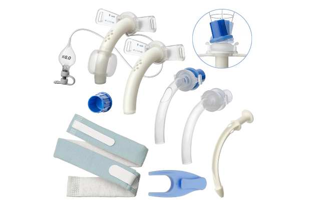
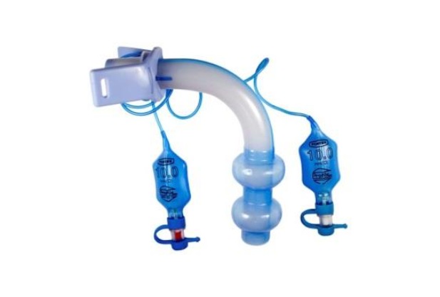
Tracheostomy tubes (cannulas) are intended to support breathing through an artificial lumen in the windpipe. These pipes differ in material, size and the presence of additional elements (cuffs, valves).

Permanent tracheostomy

Permanent tracheostomy is installed for a long time. It is placed against the background of a complete blockage of the natural respiratory tract.

This tracheostomy is indicated for patients with chronic diseases of the respiratory system and neurological disorders. It is also placed after severe neck and head injuries. Contraindications include:

  • Uncontrolled infections in the surgical area
  • Severe coagulopathies
  • High risk of rejection

The main advantage of such a tracheostomy is the possibility of long-term breathing support and facilitating care for a patient in serious condition. The main disadvantages are the risk of infection (approximately 10%), possible damage to the vocal cords and the need for constant stoma care.

Get a consultation

Temporary tracheostomy

A temporary tracheostomy is a short-term surgical opening. The doctor creates it to support breathing during recovery from acute illnesses or operations.

Indications for the installation of a temporary tracheostomy are acute conditions requiring immediate provision of airway patency (for example, laryngeal edema and tracheal stenosis). Contraindications are the anatomical features of the patient and the presence of skin infections in the area of the intended incision.

The operation is performed under local or general anesthesia. The surgeon then makes an incision on the front of the neck and creates a hole in the throat to insert the drain.

The advantages of this method include the possibility of quickly restoring respiratory functions and the relatively easy removal of the tube after the condition improves. Disadvantages include the risk of developing infections and trauma to the trachea, as well as the likelihood of scarring at the stoma site.

Get a consultation

Cuffed tracheostomy

A cuffed tracheostomy is a tube with an inflatable cuff. It provides a seal around the drain, preventing air leakage and aspiration.

Cuffed tracheostomy is used against the background of mechanical ventilation (in this case, strict maintenance of air pressure is required). Contraindications are injuries in the bronchi area (additional pressure from the cuff can aggravate the damage) and poor condition of the skin around the intended intervention site.

The main advantage is the improved ability to ventilate the lungs. In addition, this prevents aspiration of contents from the oropharynx. However, the use of a cuff tube is associated with the risk of injury to the throat (due to the pressure of the cuff) and the likelihood of developing nosocomial infections.

Get a consultation

Tracheostomy placement procedure

Fixation of a tracheostomy requires the use of special medical instruments and strict adherence to technique. The tracheostomy process looks like this:

1
Access to the throat. The surgeon makes a horizontal incision in the neck about 4-6 cm long (usually at the level of 2-4 tracheal rings). In this case, a surgical scalpel is used. To gain access to the front wall of the throat, the specialist cuts the skin, subcutaneous layer and muscle tissue. An electrocoagulator is often used to cut off tissue, which also helps stop bleeding.
2
Freeing the throat. To clearly visualize the organs, the surgeon carefully pushes away surrounding tissue and soft structures. For this, surgical clamps and special instruments (retractors) are used.
3
Creating a hole. After sufficient visualization of the bronchi, the surgeon makes an incision on one of the tracheal rings. This is usually done between the 2nd and 4th ring. In this case, a special surgical scalpel (tracheotome) is used. It allows you to accurately and accurately create a hole of the desired size
4
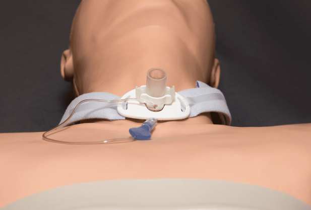
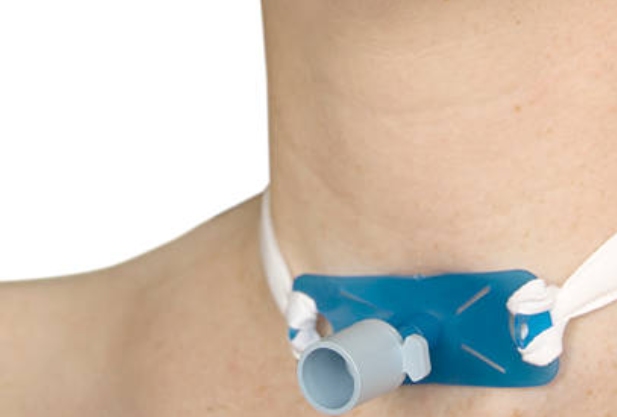
Pipe insertion. Immediately after creating the hole, a tracheostomy tube of the appropriate size is inserted into the throat. This ensures its stable position and minimizes discomfort for the patient. The drainage is fixed on the neck using special tapes or belts

To make sure that air passes freely through the drainage, auscultation (listening) of the lungs is performed. In order to visually assess the correct location of the catheter in the throat, fiberoptic bronchoscopy is used.

General information

Indications for tracheostomy surgery

Tracheostomy is prescribed when other treatment methods are ineffective. This procedure is recommended in various clinical situations associated with acute or chronic respiratory failure. It helps bypass obstructions (obstructions) or damage in the upper airway. This ensures unhindered access of air to the lungs.

The main indications are:

  • Blocked airways (this often occurs due to trauma, foreign bodies, and inflammation in the larynx or trachea)
  • Long-term use of a ventilator. This creates a need to provide air bypassing damaged or insufficiently functioning airways
  • Severe infections and diseases causing swelling or obstruction of the airways (acute laryngitis, epiglottitis)

The decision to perform a tracheostomy is based on a comprehensive assessment of the patient's condition. In addition, the doctor assesses the patient's current and potential breathing abilities. The possible risks of tracheostomy must also be taken into account.

Contraindications for surgery

There are several contraindications to tracheostomy. Here are the main ones:

  • Anatomical features (curvature of the cervical vertebrae, abnormalities of neck development) that can complicate access to the bronchi or increase the risk of damage to surrounding tissues and blood vessels
  • The presence of active infectious processes in the area of ​​the proposed surgical intervention (boils, abscesses and herpetic eruptions)
  • Coagulopathies. Bleeding disorders can lead to unnecessary bleeding during and after surgery
  • Severe general diseases. Decompensated forms of cardiac, renal and liver failure significantly increase the risk of surgical and postoperative complications

A significant contraindication is acute inflammation of the respiratory tract. For example, acute respiratory infections often worsen after surgery.

Preparation for tracheostomy

The first stage is examination. It involves passing general and biochemical blood tests, a coagulogram, a chest x-ray and, if necessary, a CT or MRI of the neck area. Further steps:

  1. Consultation with an anesthesiologist. The specialist assesses the risks associated with anesthesia and selects the appropriate type of anesthesia
  2. Preparation of the surgical field. On the eve of the operation, depilation is carried out in the place where the incision will be made. Then, in order to prevent the spread of infection, this area is thoroughly washed with antiseptic agents
  3. Preparation of tools. Immediately before the operation, surgical instruments are sterilized and placed in the required order. These include a scalpel for cutting, forceps to stop bleeding, special tissue scissors and instruments for drainage placement

8 hours before surgery, the patient should not eat or drink. On the day of surgery, the patient is usually prescribed medications to help them relax and reduce anxiety.

Possible complications

Like any surgical procedure, tracheostomy carries risks. Possible complications include:

  • Bleeding. Occurs during tracheostomy or immediately after it. The main reason is damage to the blood vessels in the incision area
  • Infection. An inflammatory process sometimes occurs in the area around the tracheostomy. If the infection is not stopped, sepsis develops
  • Pneumothorax. Air enters the space around the lungs. This causes their partial or complete collapse
  • Damage to the trachea. Wearing a pipe for a long time sometimes leads to damage to the walls of the throat and the development of scars. In some cases, stenosis develops
  • Swallowing problems. This is due to the fact that the tracheostomy puts pressure on the esophagus
  • Granulomas. They are formed in response to irritation from the catheter

To prevent the risk of complications, you must strictly follow the doctors' instructions for caring for the tracheostomy and the surrounding skin. In particular, it is necessary to regularly change bandages and monitor the cleanliness of the air that a person inhales.

Speech with tracheostomy

One of the important stages of rehabilitation of people with a tracheostomy is maintaining their speech ability. This is due to the fact that a tracheostomy makes speaking difficult: the air needed to produce sounds bypasses the vocal cords. There are ways that can help patients with tracheostomy. These include:

  • Speech restoration. The process begins with the use of special tracheostomy tubes, which allow some of the exhaled air to be directed through the vocal cords. These tubes have valves that direct air to the vocal cords when you exhale, allowing you to produce sounds
  • Using talking valves. This is a small device that attaches to the end of a tube. The valve opens when you inhale, allowing air to enter the lungs. It closes when you exhale, forcing air through the vocal cords. This is what allows the patient to speak
  • Speech rehabilitation techniques. Working with speech therapists includes breathing exercises. These techniques help control air flow and train vocal cords and articulation. The main goal of therapy is to maximize the restoration of speech functions and improve voice quality

In addition to physical rehabilitation, patients experiencing communication difficulties due to a tracheostomy should be provided with emotional and psychological support. Support from family and friends can help them adjust to new conditions and restore self-confidence.

Tracheostomy care

Proper care helps reduce the risk of complications and promotes rapid restoration of respiratory functions. The main stages of caring for a tracheostomy are as follows:

  1. Regular cleaning. To clean the skin around the tracheostomy, it is recommended to use sterile wipes or cotton swabs soaked in saline solution. To avoid causing irritation or damage, treat the skin around the tracheostomy carefully
  2. Replacing the cannula. It should be changed regularly. This is usually done by a doctor, but in some cases trained family members can perform this procedure at home. Replacement involves removing the old cannula and installing a new one. The process is performed using sterile instruments and gloves
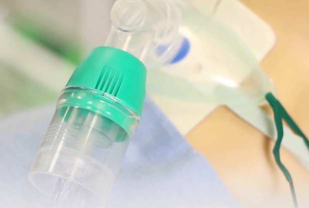
  3. Air humidification. bypasses the nose and throat. The humidity of the inhaled air is not naturally regulated in the presence of a tracheostomy tube. A humidifier can help prevent your airways from drying out and becoming irritated. If available, air enters through special humidification systems connected to the pipe
  4. Monitoring the condition of the tracheostomy. Regular examination can look for signs of infection, redness, swelling or unusual discharge. If any abnormalities are detected, you should immediately consult a doctor

If the tracheostomy has a speaking valve, its condition also requires regular monitoring and care (the principles are the same).

Recommendations after tracheostomy

People with a traeostomy should learn how to breathe correctly. This can be done using special deep breathing exercises. In this way, the risk of secretion stagnation in the lungs can be prevented.

It is very important to be aware of the signs of infection. These include redness, swelling, pain in the tracheostomy area, as well as fever, foul odor and unusual discharge.

After tracheostomy, it is necessary to change your diet (this is especially true if you have difficulty swallowing). If necessary, you should switch to specialized nutrition.

In order to monitor the condition of the tracheostomy after discharge from the hospital, the patient should regularly visit the doctor. This will help the specialist to respond to any changes in a timely manner.

Answers to popular questions

People often ask about what a tracheostomy is and what effect it has. They are also interested in caring for a tracheostomy tube and the features of living with it.

What is the difference between tracheotomy and tracheostomy?

Tracheotomy is the surgical process of creating an opening in the trachea. It allows temporary access to the airway. A tracheostomy is the placement of a special tube into this opening for long-term use.

Tracheotomy is mainly aimed at short-term solution of breathing problems. A tracheostomy provides continuous ventilation.

How often should a tracheostomy tube be changed?

The frequency of tracheostomy catheter replacement depends on its type and patient care. On average, the tracheostomy should be replaced every 1-3 months. However, if the tube becomes dirty or damaged, replacement must be done immediately. The decision about this is made by the attending physician.

Can I eat and drink with a tracheostomy tube?

Patients with a tracheostomy valve can eat and drink, but they must follow a diet. Food should be soft and easy to swallow. In some cases (for example, to adapt the diet and teach safe swallowing techniques), consultation with a speech therapist is required.

How to live with a permanent tracheostomy?

Life changes quite a lot after installing drainage. For example, difficulties arise with going outside and with hygiene procedures: in cold weather you should stay at home, and you should only take a shower wearing a special mask. Periodically (the time interval will be determined by the doctor) it will be necessary to do inhalations.

In addition, a person with a tracheostomy needs special Velcro fasteners. They are easy to put on and take off and easy to care for.
Our doctors
Sadikov
Ilya Sergeyevich
Head of the Clinic for Otorhinolaryngology, Otorhinolaryngologist
Tetzoeva
Zalina Muratovna
Head of the department of otorhinolaryngology, otorhinolaryngologist
Lapshina
Anastasia Andreevna
Otolaryngologist, audiologist
Lopatin
Andrew Stanislavovich
Chief Specialist in Otorhinolaryngology
Portnyagina
Maria Pavlovna
Audiologist-otorhinolaryngologist
Kaspranskaya
Galina Rustemovna
Otorhinolaryngologist, otoneurologist, audiologist
Budeikina
Liliya Sergeevna
Otorhinolaryngologist, phoniatrist
Erofeecheva
Irina Vladimirovna
Otolaryngologist, pediatric otolaryngologist
All specialists
2GIS Award

2GIS Award

This award is given to clinics with the highest ratings according to user ratings, a large number of requests from this site, and in the absence of critical violations.

«Good place» according to Yandex

«Good place» according to Yandex

This award is given to clinics with the highest ratings according to user ratings. It means that the place is known, loved, and definitely worth visiting.

Our doctors are laureates of the ProDoctors Award

Our doctors are laureates of the ProDoctors Award

The ProDoctors portal collected 500 thousand reviews, compiled a rating of doctors based on them and awarded the best. We are proud that our doctors are among those awarded.

Make an appointment at a convenient time on the nearest date

Price

Otorhinolaryngologist primary appointment
from 4 950 ₽
Otolaryngologist repeated appointment
from 4 950 ₽
Reception of an otorhinolaryngologist at home in Moscow within the Moscow Ring Road
from 16 300 ₽
Planned remote online consultation of an otorhinolaryngologist using telemedicine technologies

Appointment to the doctor

Fill out the form, our managers will contact you within 15 minutes
Почему К+31?
К + 31 — full-cycle multidisciplinary medical centers, including the possibility of providing medical services of European quality level.
К + 31 — are leading doctors and diagnostics using high-tech equipment from world manufacturers (Karl Storz, Olympus, Siemens, Toshiba, Bausch&Lomb, Technolas, Zeiss, Topcon).
К + 31 — is ethical. The staff of K+31 clinics maintain open relationships with patients and partners. An individual approach to each patient is the basis of our service standards.

К + 31 — is modernity. On call 24/7: call center operators will answer your questions at any time and book you an appointment with doctors. Contact us by phone, through the feedback form on the website and WhatsApp.
Our clinics
K+31 on Lobachevskogo

st. Lobachevskogo, 42/4

+7 499 999-31-31

Subway
1
11
Prospect Vernadsky Station
By a car
Lobachevsky, we pass the first barrier (security post of the City Clinical Hospital No. 31), turn right at the second barrier (security post K+31)
Parking pass
Opening hours
Mon-Fri: 08:00 – 21:00
Saturday: 09:00 – 19:00
Sunday: 09:00 – 18:00
K+31 Petrovskie Vorota

1st Kolobovsky pereulok, 4

74999993131

Subway
9
Tsvetnoy Bulvar
10
Trubnaya
By a car
Moving along Petrovsky Boulevard, turn onto st. Petrovka, right after - on the 1st Kolobovsky per. Municipal parking
Opening hours
Mon-Fri: 08:00 – 21:00
Sat-Sun: 09:00 – 19:00
K+31 West

Orshanskaya, 16/2; Ak. Pavlova, 22

74999993131

Subway
3
Molodezhnaya
By a car
Moving along Orshanskaya street, we turn to the barrier with the guard post K+31. You do not need to order a pass, they will open it for you
Opening hours
Mon-Fri: 08:00 – 21:00
Sat-Sun: 09:00 – 18:00
Didn't find the service you were looking for?