Diagnosis of benign breast formations
Modern methods for diagnosing benign neoplasms, including various biopsy options under the control of ultrasound or using stereotactic guidance, make it possible to determine the diagnosis in most cases, but even the absence of malignant growth at the time of the biopsy does not preclude its occurrence in the future.
In this regard, excision of the pathological lesion, without affecting the surrounding tissue, is the most effective treatment for this pathology.
At the present level, the removal of benign or borderline formations is carried out using the principles of oncology and plastic surgery.
Treatment of benign breast formations in K + 31
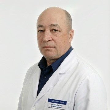
In the Department of Surgery K + 31, highly qualified oncologists-mammologists perform functionally gentle surgical interventions for any location and size of the tumor. The optimal surgical approaches in this case are inframammary and periareolar. These areas are less prone to the formation of visible scars and, at the same time, provide access to almost any part of the mammary gland with modern retractors and good lighting. By the way, they also make the cut length shorter and provide optimal healing. When the tumor is localized high in the upper-outer quadrant or accessory lobule, transaxillary access is also used.
K + 31 is equipped with modern ultrasound machines that allow for intraoperative localization of non-palpable formations and control during their removal. The advantage of the method is that it allows the doctor to see all areas of the mammary gland, including hard-to-reach areas that are located near the chest wall, as well as non-palpable small tumors. This reduces the risk of excess trauma to the breast tissue during surgery and shortens the time of postoperative rehabilitation.
Timely removal of benign breast tumors can not only reveal hidden forms of breast cancer, but also prevent its development in proliferation zones.

