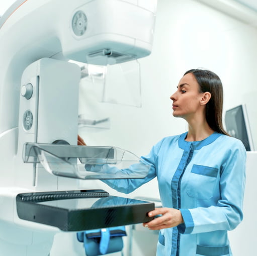
Mammography is a non-invasive method for examining the mammary glands. Women over 40 need to undergo this procedure annually. It is also indicated in cases where there is a seal in one or both mammary glands, uncharacteristic discharge from the nipples, changes in the skin on the chest, etc.
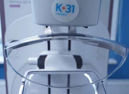
In the K+31 medical center, modern Senographe Pristina™ 3D equipment is used to conduct this type of examination. This ensures the informativeness of the results with minimal exposure of the patient. You can make an appointment for a mammogram by phone or through the feedback form on the website.

specialists

mammographs

techniques
Diagnosis of diseases
Mammography is a fast, painless and accurate method of functional diagnostics used in mammology. The study is carried out mainly to detect benign and malignant tumors. Unlike ultrasound, a mammogram allows not only to identify a neoplasm, but to see its size and determine its nature. With this method, you can detect the tumor at an early stage and start treatment for breast cancer as soon as possible.
Many women are afraid that it is harmful to do a mammogram, because X-rays are used in the study. Of particular concern is the presence of existing tumors - would mammography be dangerous in this case? The answer is more than unequivocal: the dose of X-rays obtained during examinations on the latest generation of mammographs, which K + 31 clinics are equipped with, does not cause any concern at all. In fact, it is lower than with radiography.
As for the presence of neoplasms, mammography is used to monitor their activity and growth. Before prescribing a study, the attending physician will necessarily take into account all risk factors and direct him to the study that will be informative in a particular case.
Types of mammography
Mammography is the most accurate research method that allows you to accurately identify pathological changes in the breast and take prompt measures to eliminate them. There are several types of mammography:
- Digital mammography
- Ultrasound
- Optical mammography
- MRI
Digital mammography
It replaced the traditional analog one (using x-rays). Differs in image accuracy, fast procedure and high information content. Images captured on the monitor can be saved on information carriers for further research and control over the dynamics of changes.
Ultrasound
Ultrasound diagnostics of the mammary glands is mainly performed for women under 40 years of age. Also, due to the safety of the method, it is used in women during pregnancy and lactation. Ultrasound is effective, but in some cases may give incorrect information. Most often, it is used as an additional study when it is difficult to make a diagnosis.
Optical mammography
Assumes the use of laser radiation. A picture with optical mammography is performed in two projections. Allows you to accurately determine the presence or absence of benign and malignant neoplasms. It is prescribed for women under 30 years old, and also to monitor the dynamics of changes in formations, if any.
MRI
MRI of the mammary glands is the most accurate and informative research method. It is completely safe because it does not involve the use x-ray or ionizing radiation.
The choice of method for examining the mammary glands is determined by the attending physician. It depends on various factors (age, medical history, presence of complaints). Reception of patients in the medical center K+31 is carried out by experienced and highly qualified doctors. You can sign up for a consultation by phone or leave a request on the website.
Indications and contraindications for breast mammography
First of all, mammography is indicated for women over 40 for preventive purposes, since it is at this age that the risk of developing cancer increases. It is recommended for women under 35 if they are at risk (hereditary predisposition to oncology, early menstruation, history of abortion, infertility, etc.).
Pros of digital mammography
Also, this type of examination is prescribed in cases where the patient has:
- Uncharacteristic chest pain
- Induration in the soft tissues of the breast
- Nipple discharge
- Breast engorgement
- Diseases of the endocrine system (thyroid gland), etc.
It is also necessary to undergo mammography after the treatment to monitor the condition and effectiveness of therapy.
Despite the fact that digital mammography is a safe procedure, it still involves a certain amount of radiation exposure. This means that she also has minimal contraindications:
- Pregnancy and lactation
- Presence of breast implants
- Injury to the skin and soft tissues of the chest

Book a mammogram
Our doctors

This award is given to clinics with the highest ratings according to user ratings, a large number of requests from this site, and in the absence of critical violations.

This award is given to clinics with the highest ratings according to user ratings. It means that the place is known, loved, and definitely worth visiting.

The ProDoctors portal collected 500 thousand reviews, compiled a rating of doctors based on them and awarded the best. We are proud that our doctors are among those awarded.
Comprehensive programs
Early diagnosis is the key to a long and healthy life!
Regular digital mammography is the only method of early detection of breast cancer with proven effectiveness.

Preparing for a mammogram
A referral for examination by a mammologist is most often given by a therapist or gynecologist. If there is a suspicion of the development of a malignant neoplasm in the breast, the patient may be additionally prescribed other methods of laboratory diagnostics.
The information content of the procedure performed largely depends on the correctness of the preparation. To prepare for a mammogram, the patient needs to know the following rules:
- The date of the study must be planned in advance. The most optimal time is 5-10 days of the cycle
- The date of the study must be planned in advance. The most optimal time is 5-10 days of the cycle
- Immediately before a mammogram, the use of deodorant is undesirable (the equipment may perceive them as calcifications)
It is also important to inform the specialist in advance about a history of surgery or suspected pregnancy.
Price
Answers to popular questions
When is a mammogram prescribed?
Digital mammography is a more advanced form of the already familiar breast x-ray. This research method is safer, more informative and convenient. This procedure is recommended not only for diagnosing diseases. It is also suitable for routine examination of women over 35 years of age. Before this age, ultrasound is considered more informative. The need for digital mammography is determined by the attending physician. The choice of medical manipulation depends on the patient’s age, health status, and the presence of concomitant diseases.
To diagnose pathological changes, mammography is prescribed for the following symptoms:
- Painful sensations in the breast, changes in its shape.
- Swelling of the mammary glands.
- Changing the shape of the areolas.
- Uncharacteristic discharge from the nipples.
- Redness of the skin on the chest.
- Recession of nipples, etc.
If you have any alarming symptoms, you should consult a mammologist as soon as possible. The specialists of the K+31 clinic find an individual approach to each patient, which guarantees a complete examination and prompt assistance in any situation.
How is mammography performed?
The digital mammography procedure is recommended to be performed on days 6-12 of the cycle. But this is not a prerequisite. Information content depends not only on the experience of the doctor and the characteristics of the mammograph. It is advisable not to move during the manipulation.
Mammography is performed as follows:
- The woman removes all clothing above the waist.
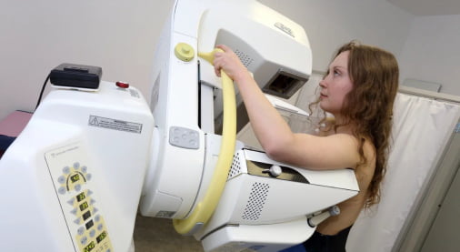
- The mammary gland is placed on the mammograph platform. The plate located on the other side presses it slightly. This is necessary so that the breasts are thinner and more uniform.
- The patient needs to hold her breath while the doctor takes a photo of the chest in a direct and oblique projection.
- The same actions are carried out with the second mammary gland.
After the mammogram, the woman gets dressed. The doctor takes pictures and records the results. The conclusion contains information about the homogeneity of breast tissue, the condition of the milk ducts, the presence of neoplasms and pathological foci.
What does mammography show?
Digital mammography allows you to diagnose various changes in the breast. Mainly malignant tumors. In addition, computed tomography of the breast allows you to diagnose the following benign tumors:
- Mastopathy (benign pathology resulting from hormonal imbalance).
- Cyst (a cavity filled with intracystic fluid).
- Calcifications (calcium salts concentrated in the mammary gland. Their presence indicates a tumor process).
- Fibroadenoma (benign tumor-like formation formed from healthy cells).
How are mammography results interpreted?
The BI-RADS scale is traditionally used to interpret mammography. The results of the study are interpreted using 6 categories:
- BI-RADS-0. There is a possibility of deviation from the norm, but the result is not informative enough to make a diagnosis.
- BI-RADS-1. There are no anomalies, the condition of the mammary glands is within normal limits.
- BI-RADS-2. A benign neoplasm was detected.
- BI-RADS-3. A benign formation has been identified, which requires observation by an oncologist until a final diagnosis is made.
- BI-RADS-4. Suspicion of malignancy. Additional laboratory testing (biopsy) is required.
- BI-RADS-5. A malignant tumor has been discovered. Extensive examination is necessary.
- BI-RADS-6. The diagnosis of cancer has been confirmed. The patient needs prompt treatment.
At the K+31 medical center, consultations are carried out by qualified specialists using modern and precise equipment. This allows you to effectively carry out all the necessary research. You can make an appointment with a mammologist at the K+31 clinic using the specified phone number or sign up for a consultation on the website.
The benefits of digital mammography
For diagnostic examinations of the mammary glands, specialists increasingly prefer digital mammography. This is due to the undeniable advantages that this procedure has:
- Possibility of quickly displaying examination results on the monitor.
- Clear image of the condition of the mammary glands, regardless of breast size.
- Quickness of the procedure.
- High accuracy of results.
- Reduced radiation dose compared to conventional radiography.
- Probability of diagnosing cancer at the earliest stages.
Other types of breast examination
In addition to digital mammography, other methods can be used to examine the mammary glands and diagnose various diseases. The most optimal method is selected by the doctor in each specific case, taking into account many factors (age, symptoms, contraindications, etc.).
Ultrasonography. A safe procedure that is suitable for women of any age. Indicated for examination in case of inflammatory processes, uncharacteristic discharge from the nipples, menstrual irregularities, control of previously diagnosed cysts, as well as after surgery.
MRI. It is not a replacement for ultrasound or mammography, but only complements existing studies. Used as a screening for patients who are at risk due to age or heredity. Helps determine the size of malignant tumors and the presence of metastases. Suitable for examining lymph nodes. An important advantage of this method is complete safety for pregnant women and very young patients. It is also suitable for women with breast implants.
Ductography of the milk ducts. A method of radiography using a contrast agent. It is a type of mammography. The main advantage of the method is a complete examination of the ducts and the ability to diagnose any pathological changes that may occur in them. Thanks to it, you can clearly determine where the tumor is located.
Other types of breast examination also include visual examination, palpation, biopsy and various cytological studies. The choice of necessary manipulations remains with the doctor, taking into account all the nuances and indications.
