Computed tomography
(CT)
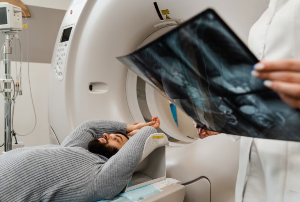
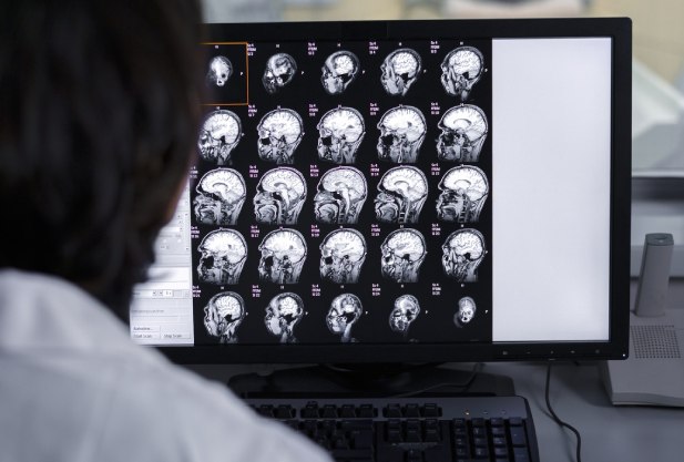
Computer tomography CT (MSCT) is a diagnostic procedure based on the principle of penetration of X-rays through the tissues of the human body. Unlike classical radiography, CT involves taking multiple images at once, obtaining "slices" and then composing them using software. Layer-by-layer study of the structures of blood vessels, bone tissue and organs allows you to get a high-quality digital image and a conclusion from a radiologist.
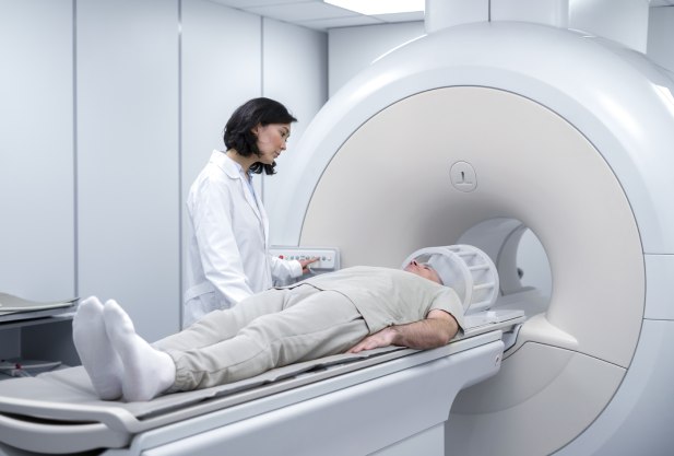
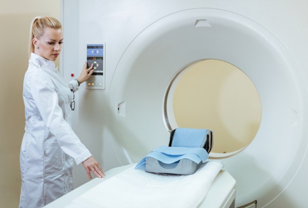
The K+31 clinics are equipped with modern computer tomographs, which allow us to provide:
- Fast scanning with minimal patient exposure.
- Obtaining high-precision images.
- High level of patient comfort and safety.
- The examination result on the same day.
Advantages of CT scan in K+31
All routine studies with dynamic contrast: brain, soft tissues of the neck, paranasal sinuses, facial part of the skull, any part of the spine, any joint, organs of the chest cavity, organs of the abdominal cavity, organs of the pelvic organs, enterography.
Specialized studies: heart, coronary angiography, virtual colonoscopy, aortic angiography, brachiocephalic angiography, limb angiography, perfusion of any organ, brain, liver, pancreas
Dual-energy CT: pulmonary artery angiography, determination of kidney stone composition, construction of iodine maps for small lesions in parenchymatous organs
CT-guided biopsy: lung, liver and other organs
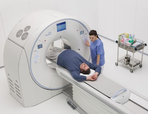
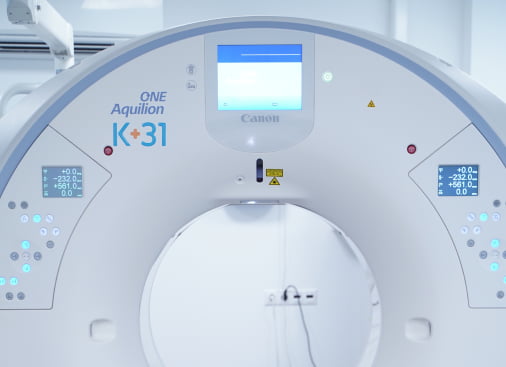
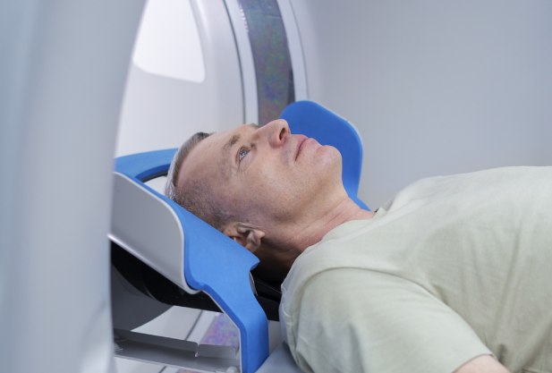
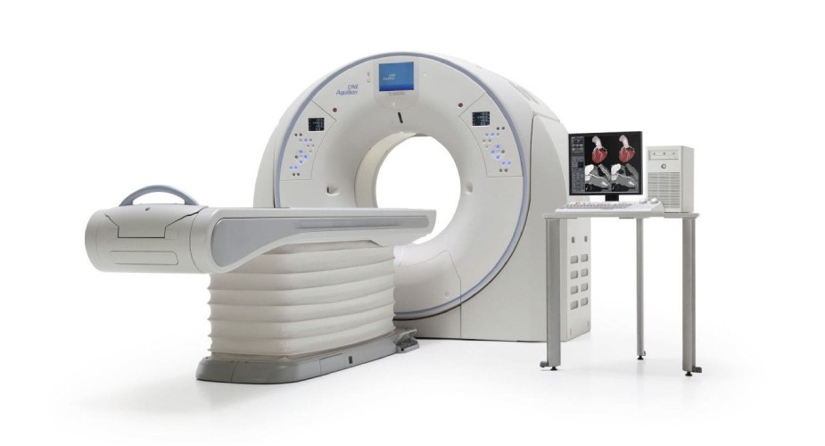
The Aquilion One Genesis Edition CT scanner used in K+31 clinics is one of the best in the world. 640 slices is the maximum value available today in diagnostic practice.
Features:
- Whole Body Scan
- 320-row detector = 640 slices per 1 gantry rotation
- Best spatial resolution performance
- Minimum radiation dose requirements
- Gantry aperture diameter - 780 mm
- Load capacity on long table - up to 315 kg
Popular services
- CT enterography
- Computed tomography of the abdominal cavity and retroperitoneal space
- Spiral computed tomography of the pelvic organs
- Computed tomography of the chest organs
- Spiral CT scan of the neck
- Computed tomography of the orbit
- Computed tomography of the jaw
- Computer tomography of the brain
- Computed tomography of the pelvic bones
CT enterography
CT enterography or computed tomography of the intestine is a modern method of radiation diagnostics of diseases and pathologies of the small intestine. The essence of the procedure is to fill it with a neutral medium to straighten the lumen of the intestine and achieve the necessary contrast of the walls. If it is necessary to obtain high-contrast visualization of the mucous membrane and feeding vessels, then diagnostics are carried out with intravenous bolus contrast.
CT enterography allows you to assess the condition of the walls, mucous membranes and feeding vessels in the intestine much better and more accurately than any other procedure. It allows you to get information about pathologies not only of the small intestine, but also of other organs of the small pelvis, abdominal cavity and retroperitoneal space. During preliminary preparation for diagnostics, the patient's intestines are cleaned and stretched with liquid contents, which allows you to identify diseases and pathologies at an early stage, starting their treatment in a timely manner. During the procedure, the doctor can assess the condition of the mucous, muscular and serous membranes of the small intestine, the nature of the changes in which accurately indicates a particular disease. Polyethylene glycol preparations used in diagnostics are absolutely safe for human health.

Appoint to CT
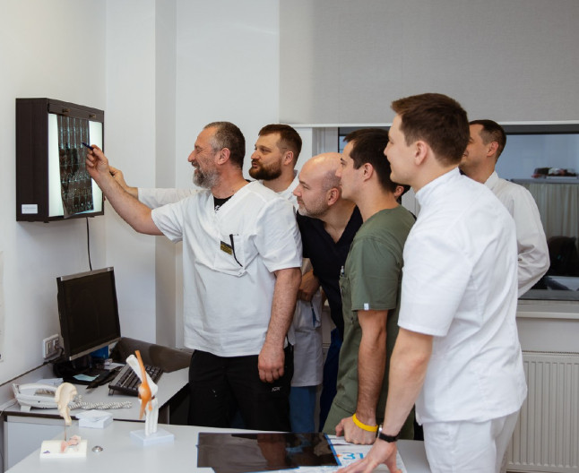
Our doctors


This award is given to clinics with the highest ratings according to user ratings, a large number of requests from this site, and in the absence of critical violations.

This award is given to clinics with the highest ratings according to user ratings. It means that the place is known, loved, and definitely worth visiting.

The ProDoctors portal collected 500 thousand reviews, compiled a rating of doctors based on them and awarded the best. We are proud that our doctors are among those awarded.
Price
Helpful information
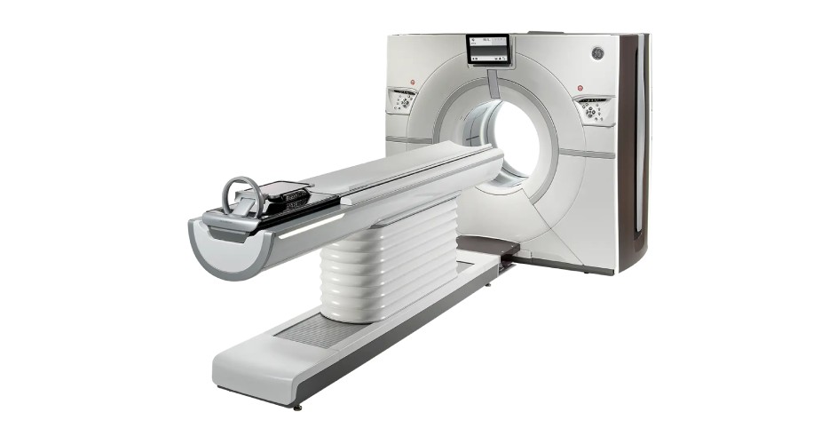
What are the technical characteristics of the tomograph in K+31
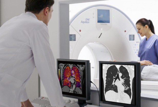
Aquilion One Genesis Edition computed tomograph, used in K+31 clinics, one of the best in the world. 640 slices are the maximum values available in diagnostic practice today. Thanks to numerous clinical research and breakthrough technological developments have managed to achieve the clearest spatial permitting and conducting research with a minimum radiation dose.
The CT has a built-in AiCE processor, which operates on the basis of a neural network for reconstructing computed tomography images. This method is characterized by deep learning and allows AiCE to make high-quality work as quickly as possible. CT images with precise detail and minimal noise.
Features of CT scanning in K+31
- Weak noise.
- Clear image.
- High contrast resolution.
- Patient-centric imaging solutions
- Clearness of the content of complex protocols.
- Consistency in providing quality results.
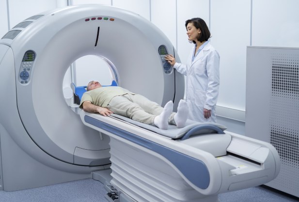
- With one rotation of the detector (fractions of a second), high-quality images are obtained in both simple and complex examination cases: the heart, the chest of a newborn, the ankle joint or foot.
Additional conveniences for the patient in K+31
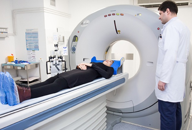
The redesigned, expanding CT scanner portal has a wide open area to accommodate patients. A large space gives a calming effect and helps a person relax before the study.
The design allows staff to easily assist patients with injuries and during interventional procedures from both the anterior and posterior CT scans.
In computed tomography at K+31 it is possible to avoid the usual scan planning procedures, since the Aquilion ONE CT laser collimation allows you to enter the field of view and scanning range directly on the portal.
Types of CT scans performed at K+31
To examine the spine and hip joints, the diagnostic center provides the opportunity to undergo 3D densitometry. To diagnose the abdominal cavity, a virtual colonoscopy is prescribed. To identify pathologies of blood vessels, veins, and arteries, CT angiography using iodine-containing contrast is recommended. Contrast is also used in oncology to detect metastases.
Be examined by us if you have been prescribed any of the following types of computed tomography:
- CT scan of the brain
- MSCT angiography of cerebral vessels with intravenous bolus contrast
- CT scan of the eye orbits
- CT scan of the chest
- CT angiography of the thoracic and abdominal aorta
- CT scan of the lungs
- CT scan of the abdominal cavity and retroperitoneum
- CT scan of the pelvic organs
- CT coccyx
- CT scan of different parts of the spine: cervical spine, thoracic spine, lumbosacral spine
- CT scan of the lower extremities (knee, ankle, thigh and lower leg)
- CT scan of paranasal sinuses, temporomandibular joint
- CT scan of the larynx
- CT scan of the kidneys and adrenal glands
- CT scan of the urinary system: bladder, ureters
- CT scan of the pelvic bones
- CT scan of the elbow and shoulder joint
- CT scan of the wrist joint
- CT scan with contrast
If necessary, our diagnostic center has machines for magnetic resonance imaging, ultrasound and other examinations. Sign up for a CT scan or call the phone number listed on the website.
Is computed tomography harmful to health?
Obviously, like any other radiology procedure involving the use of x-rays, CT implies a certain dose of radiation exposure to the patient's body. The doctor also understands this, so the appointment CT scan is always done based on the diagnostician required to make a diagnosis. information and the patient's condition, can reliably determine the need and frequency of prescribing a particular procedure.
Is CT examination harmful to health? Yes, of course, radiation exposure has a certain effect on the body, but in acceptable doses. At the same time, modern equipment for computed tomography is also manufactured based on the principle of minimizing radiation exposure. New technological tomographs have emitters reduced by several times in comparison with the previous generation of x-ray beam power, and the time of its impact on the body is as limited as possible.
Accurate visualization of disease progression with CT
Valuable additional information in the form of images obtained using computed tomography, and added to morphological studies contributes to the optimization of disease surveillance and treatment methods. The most complete perfusion maps are indispensable in the diagnosis and therapeutic response in tumors or strokes. Dynamic CT scans performed on a tomograph will be able to find out the causes of immobility or pain in the joints.
General recommendations and procedure for performing CT examinations
- Coming to a CT examination should be done with a minimum of cosmetics.
- Temporary tattoos in the study area must be removed.
- Underwear (t-shirts, nightgowns, etc.) should be free of rhinestones, no appliqués.
- Do not eat 2 hours before the CT scan with contrast.
- You must bring previous images and descriptions of ultrasound, CT, MRI with you.
- Time to conduct a CT scan - from 15 to 40 minutes.
When is preparation for CT needed?
Some types of computed tomography (CT) scans require special preparation for the patient. If a CT scan with a contrast agent is prescribed, it is necessary to make sure that there are no contraindications. To do this, the patient takes a biochemical blood test for creatinine, urea, glucose, an allergen for a contrast agent (iodine).
For CT scans without contrast agent, no preparation is necessary in most cases. You can find out more about the preparation in the contact center of the clinic, with a doctor or in your personal account.
Reviews 10