Patent received on the basis of K + 31 Petrovsky Gates
Diseases of the temporomandibular joints (TMJ) are common and occur in 25-65% of the population. TMJ dysfunction is the most common condition among this group of disorders and accounts for about 80-90% of the total number of patients. Most often, the lesion is characterized by the presence of pain in the joint, extending to the area of the eyes and temple, which can be aggravated with wide opening of the mouth and lead to reflex spasm of the masticatory muscles. Changes in TMJ can be assessed using different research methods, but the most informative is magnetic resonance imaging (MRI).
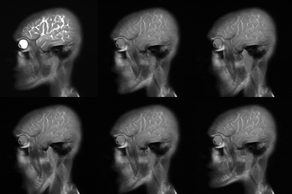
At the end of 2018, a patent was obtained on the basis of AO K + 31 Petrovsky Gates on the basis of real-time MRI dysfunction assessment using MRI. The technique allows you to assess the ratio of bone structures in the joint during the natural opening of the mouth. In addition, the position of the intraarticular disc is evaluated, the displacement of which is a critical criterion for dysfunction.

Magnetic resonance imaging is performed on a Siemens Essenza apparatus with a magnetic field induction of 1.5 T. The study does not require special patient preparation. The procedure is carried out using a standard matrix 16-channel transceiver coil for the head. The study protocol is divided into three parts: 1. in the position of natural mouth occlusion; 2. in the open mouth position using a bite retractor; 3. A series of dynamic images in real time.
Static images represent the anatomy of the joint at the end points of movement and do not give a complete picture of the displacement of the intraarticular disk. Real-time MRI clearly determines the configuration of the intraarticular disk with obtaining objective data on its movement at all stages of the active movement of the jaw, which allows us to make a correct conclusion about the degree of joint dysfunction.
The inventors are a radiologist at the Department of Radiation Diagnostics K + 31 Dushkova Daria Vladimirovna, her scientific adviser is Ph.D. senior scientist SPC MR Vasiliev Yuri Alexandrovich and a consultant on the osteoarticular system K + 31 Ph.D. Vasilieva Julia Nikolaevna.

